
Authors:
Becceneri, Amanda B. [1] ; Fuzer, Angelina M. [1] ; Plutin, Ana M. [2] ; Batista, Alzir A. [3] ; Lelievre, Sophie A. [4] ; Cominetti, Marcia R. [1]
Abstract:
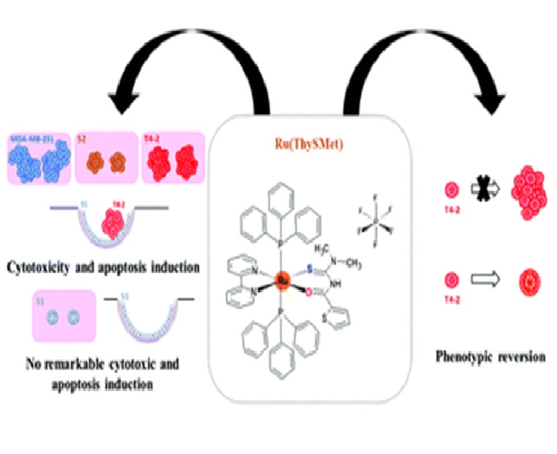
Many studies have revealed the advantages of using three-dimensional (3D) culture over traditional twodimensional (2D) monolayer techniques. The 3D cell culture models represent biologically relevant approaches to better mimic the tumor organization observed in vivo. These models have high potential for anticancer drug development and screening, for which challenges include tumor cell resistance to treatment and the risk of toxicity for patients. Many anticancer drug candidates do not reach clinical trials as they fail preclinical tests in vivo, although they were promising in 2D cultures. Models from 3D cell cultures are proposed as intermediate screening filters between 2D and in vivo assays. It has been suggested that ruthenium complexes have great potential for breast cancer treatment. Here, we tested the trans{[}Ru(PPh3)(2)(N,N-dimethyl-N-thiophenylthioureato-k(2)O,S)(bipy)]P F6 complex in breast cancer cell lines using different 3D culture techniques, including the embedded, `on top’ and disease-on-a-chip (DOC) models that reproduce physical aspects of the tumor microenvironment. The complex had pronounced but distinct cytotoxic effects on tumors cultured in collagen I of appropriate stiffness for the disease stage; the extent of induced apoptosis depends on the preinvasive (S2 cells) and invasive (T4-2 and MDA-MB-231 cells) nature of triple-negative breast cancer models. Remarkably, in the DOC model, which simulates breast ductal architecture, the complex was cytotoxic for T4-2 tumors but had no remarkable effect on the differentiated luminal epithelial S1 cells, demonstrating selectivity against cancer cells. In addition, a lower concentration of the complex abrogated the malignant phenotype of the T4-2 cells with reduction of EGFR, p50 NF kappa B, and beta 1-integrin expression. To the best of our knowledge, this work uniquely demonstrates the effects of a ruthenium complex on the induction of apoptosis and on the phenotypic reversion of tumor cells in 3D cultures. These results warrant moving to in vivo evaluation of this ruthenium complex.
1 Department of Gerontology, Federal University of São Carlos, Rod. Washington Luís, Km 235, São Carlos, São Paulo, Brazil
2 Facultad de Química, Universidad de la Habana, Zapata s/n entre G y Carlitos Aguirre, Habana, Cuba
3 Department of Chemistry, Federal University of São Carlos, Rod. Washington Luís, Km 235, São Carlos, São Paulo, Brazil
4 Department of Basic Medical Sciences and Center for Cancer Research, Purdue University, 625 Harrison Street, West Lafayette, Indiana, USA
Link to article: https://pubs.rsc.org/en/content/articlelanding/2020/QI/D0QI00502A#!divAbstract